The lung is one of the most essential organs in humans, but it likewise hosts some dreadful conditions and illness. Atelectasis is among the conditions where the air sacs (alveoli) in the lungs deflate unusually. In this case, the result is a total or partial collapse of the lung(s). Typically, this issue arises after surgery. The grave implications of atelectasis are immediately evident as it hinders the quantity of oxygen within the body.
Bibasilar atelectasis affects the left and right bottom parts of the lungs. This short article will explain in detail its causes, symptoms in addition to treatments so that you can better deal with or avoid this condition.
What Is Bibasilar Atelectasis?
Basilar atelectasis is the name offered to the condition, where either a part of the lung or the whole lung collapses due to a barrier. This condition impacts both the left and right lungs. It can be chronic or acute, preventing the breathing exchange of oxygen and dioxide. Due to the fact that of the collapsing of the lungs, another condition called bibasilar scarring happens because part of the lung. Another derivation of this condition is bibasilar sub-segmental atelectasis, which leads to the compression or disintegration of a part of the lung distal to blocked segmental bronchus.

What Happens in Basilar Atelectasis?
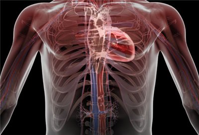
Understanding about the lung is important when finding out about basilar atelectasis. A body has a set of lungs, located on the left and right sides of the chest. Contrary to good sense, both lungs are not similar; in truth, the left lung has only two partitions, compared to the three of the right lung. Both lungs include small air sacs frequently described as alveoli. These sacs are filled with blood vessels, and are constantly involved in gas exchange.
When the bottom of the lung (right or left) collapses because of an obstruction, it causes gas exchange to halt. This condition is known as basilar atelectasis.
Bibasilar Subsegmental Atelectasis
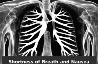
Subsegmental atelectasis is specified as the decrease in the volume of the lung due to the impediment of the little or subsegmental bronchus. The condition appears as direct opacity in a chest radiograph. People struggling with respiratory tract infection, hypoventilation, and pulmonary embolism are usually affected with subsegmental atelectasis.
Symptoms of Bibasilar Subsegmental Atelectasis
- Low fever
- Coughing
- Shallow, quick breathing
- Dyspnea (problem in breathing).
What Happen If You Have Mild Bibasilar Atelectasis?
Health conditions and injury to the lungs can also bring upon bibasilar atelectasis. However, mild bibasilar atelectasis do not need treatment while more severe cases need surgery. Anyway, iytmed.com team strongly recommends you contact your doctor if you have any of bibasilar atelectasis symptoms mentioned below.
In medicine, there are a number of variations of the disease as basilar subsegmental atelectasis, mild bibasilar atelectasis, segmental atelectasis, etc.
Symptoms of Bibasilar Atelectasis
Typically, the symptoms of atelectasis are tough to select as they are similar to those of other illness or conditions and can be mistaken to be signs of something else. The symptoms of atelectasis are normally:
- Difficulty in breathing
- Fast or shallow breathing (just like breathing after any difficult exercise)
- Coughing
- Low fever
Complications
The complications arising from atelectasis can be quite disturbing. They include:
- Low blood oxygen (hypoxemia): this happens due to the fact that atelectasis hampers the amount of oxygen the alveoli receive.
- Pneumonia: among the factors for atelectasis is plugs of mucus. The mucus within a lung impacted by atelectasis is an attractive hotbed for various bacterial infections. This can result in pneumonia.
- Lung scarring: after a lung has collapsed, it needs to be re-inflated. Re-inflation, nevertheless, at times, cannot heal all the damage and scarring, which in turn may cause bronchiectasis.
- Breathing failure: Atelectasis covering a small area is usually treatable. Having atelectasis as an adult is relatively better, in babies (as well as grownups with lung diseases), it can prove to be lethal if it covers enough area.
See your doctor right away if you experience any problems breathing.
Causes of Bibasilar Atelectasis
Atelectasis, generally, results from obstructed air passages. Since, anesthesia obstructs the routine airflow-pattern of the lungs, causing a change in absorption of gases. This, in turn, plays a part in the collapsing of the alveoli in the lungs. The other reasons for an obstruction in the lungs (obstructive atelectasis) include:
- Foreign body: any foreign item, which gets inhaled instead of ingested, can cause a collapse of the lungs.
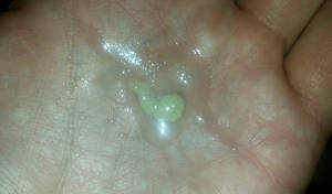
- Mucus plug: during surgery, the drugs injected into the patient cause the lungs to have actually reduced inflation, triggering the mucus to gather in the respiratory tracts. A regular, medical practice of suction of the lungs helps suppress this situation; however, the mucus might keep developing later on. This is one of the factors doctors recommend deep breathing exercises during recovery. Mucus plugs are also discovered in people suffering from cystic fibrosis, and asthma.
- Embolism: experiencing severe physical trauma can cause blood to spill out inside the lungs, which, often can’t be coughed out.
- Narrowing of significant airways from disease: diseases, like tuberculosis and fungal infection, can give rise to scarring in the lungs which restricts the airways.
- Growth in a significant respiratory tract: given that a growth is currently an obstruction, it can cause atelectasis.
Possible Causes of Non-obstructive Atelectasis Include:
- Injury: Any major injury can cause the victim to have lowered breathing due to the pain exhibited. This can lead to compression of the lungs, triggering atelectasis.
- Pneumothorax. Air from lungs enters into the space in between lungs and chest wall, indirectly triggering a little or whole of the lung to obtain damaged.
Also read: Pain in Lungs when Breathing
Diagnosis of Bibasilar Atelectasis
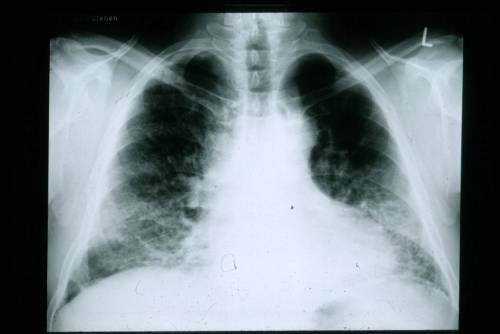
The most common method to find atelectasis is an x-ray as it can show any sort of obstruction within the lungs. Other tests include CT scans, ultrasounds, oximetry tests and bronchoscopy.
Treatment for Bibasilar Atelectasis
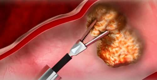
The treatment is highly based on the condition. If only a small area has been impacted, no treatment might be needed. If there is an outside obstruction, like a tumor, the treatment concentrates on eliminating or shrinking the obstruction.
The majority of doctors encourage coughing, deep breathing exercises and postural drain after surgery to assist re-inflate the lungs.
Medicines, like Foradil (given to patients with bronchitis) expand the bronchial tubes, making it simpler to breathe. Acetadote is used to make the mucus thin and easier to cough up.
It’s important to know that cigarette smoking can increase mucus production and damage the wall. It is basically important to stop smoking cigarettes in order to avoid the condition to get worse and prevent it from taking place in the first location.