
Dermatologic symptoms are seen in 25 % of clients with sarcoidosis. They usually accompany systemic involvement, but in some cases they might be the only symptoms of the illness. Sarcoidosis is defined by noncaseating epithelioid granulomas that may influence any organ system.
Although skin specialist described the very first case in 1869, the etiology of the condition is still unidentified. The illness most typically includes granuloma development in the lungs, with 90-95 % of clients having some lung participation. Other frequently included organ systems include the lymph nodes (particularly the intrathoracic nodes), the skin, the eyes, the liver, the heart, and the anxious, musculoskeletal, kidney, and endocrine systems.
Physical Exam for Sarcoidosis Skin
Cutaneous participation is classified as either “particular” or “nonspecific”. Histopathologically, certain sores materialize as noncaseating granulomas, whereas nonspecific sores do not expose granulomas on histopathologic examination. Erythema nodosum (EN) (shown below) is the main nonspecific cutaneous condition. Lupus pernio, maculopapular, nodular, scar, plaque, angiolupoid, ichthyosiform, lichenoid, annular, verrucous, psoriasiform, and ulcerative lesions and subcutaneous nodules are examples of certain cutaneous condition.
Blau syndrome and early-onset sarcoidosis represent familial and sporadic types of pediatric granulomatous autoinflammatory illness caused by anomalies in the NOD2 gene. Clinical onset is normally early in life, with noncaseating granulomas affecting the skin, joints, and uveal system.
Types of Sarcoidosis Skin Rash
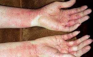
Erythema nodosum
EN is a hypersensitivity reaction resulting from exposure to a range of transmittable representatives (especially current streptococcal infection), drugs (consisting of contraceptive pills), or systemic inflammatory disorders (eg, sarcoidosis, inflammatory bowel illness). EN is usually an intense, self-limiting procedure and rarely needs treatment. Recurrences are uncommon. Tender, erythematous blemishes are usually present on the extremities, a lot of commonly on the anterior surface area of the tibia. Fever, arthralgia, and despair may take place. EN is more typical in European, specifically Scandinavian, women of childbearing age than in other people.
Loefgren syndrome
Löfgren syndrome is classically referred to as a triad of EN, polyarthritis, and hilar adenopathy. The adenopathy may be unilateral or bilateral hilar and/or ideal paratracheal lymphadenopathy. Other signs consist of anterior uveitis, fever, ankle periarthritis, arthralgias, and lung participation.
Löfgren syndrome is normally a severe illness with an excellent prognosis, usually solving spontaneously from 6-8 weeks to approximately 2 years after beginning. Pulmonologists, eye doctors, and rheumatologists often define this syndrome differently, describing differing combinations of arthritis, arthralgia, uveitis, EN, hilar adenopathy, and/or other scientific findings.
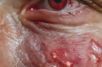
Lupus pernio
Lupus pernio, very first explained by Besnier in 1889, is a striking symptom of sarcoidal skin lesions. Lupus pernio is characterized by red to purple or violaceous, indurated plaques and blemishes that generally affect the nose, cheeks, ears, and lips, but it can appear on the dorsa of the hands, and on the fingers, toes, and forehead.
Lupus pernio is normally more typical in black women with long-standing systemic, typically lung, sarcoidosis than in other individuals. It is also frequently seen with persistent uveitis and bone cysts. The course is normally persistent, might be more recalcitrant to treatment, and may result in serious cosmetic disfigurement. Lupus pernio, particularly including the nasal rim, has actually been associated with granulomatous involvement of the upper breathing system (50 %) and lungs (75 %). In addition, it is associated with a higher frequency of ocular involvement, bone cyst formation, and lymphadenopathy or organomegaly.
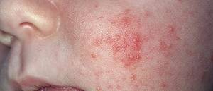
Macular sarcoidosis
Macular or papular sarcoidosis is the most common lesion seen in cutaneous sarcoidosis, especially in black ladies. Granulomatous acne rosacea may mimic sarcoidosis medically and histopathologically. Normally, sores are asymptomatic, red-brown macules and papules typically including the face, the periorbital locations, the nasolabial folds, and/or the extensor surfaces. Lesions typically fix without scarring, although scarring might take place. These lesions may likewise occur in severe sarcoidosis
Making use of dermoscopy to help in the medical diagnosis of macular and plaque-type sarcoidosis has actually been reported, with findings of “clear yellow to orange globular-like or structureless locations associated with linear vessels” and being related to granulomatous skin disease, including cutaneous sarcoid. [4] Widespread atrophic lesions with elastolysis have actually been reported, and widespread lichenoid sores might look like erythroderma.
Plaque sarcoidosis
Plaque sarcoidosis is defined by round to oval, red-brown to purple, infiltrated plaques; the center of the plaque might be atrophic (see the image listed below). Some plaques might even appear scaly and can be confused with sores of psoriasis or lichen planus. Dermoscopy may help in the clinical diagnosis, as noted above.
The sores most frequently take place on the extremities, face, scalp, back, and buttocks, and they might have an annular look (see the image listed below). The distribution is normally in proportion. Angiolupoid sarcoidosis is a subtype that has a comparable look but has huge, telangiectatic vessels in addition to the characteristics pointed out above. This kind of cutaneous participation is typically chronic; most patients have the illness for more than 2 years. Lesions can heal with scarring, and, if plaques involve the scalp, they may lead to alopecia. Patients with plaque sores normally have more severe systemic participation.
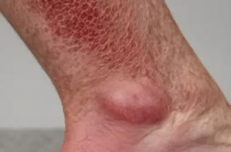
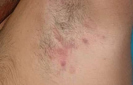
Subcutaneous nodular sarcoidosis
Subcutaneous nodular sarcoidosis is also called Darier-Roussy sarcoidosis. Sores are normally nontender, firm, oval, flesh-colored or violaceous nodules that are 0.5-2 cm in diameter. They are typically discovered on the extremities or trunk. These sores usually appear in the start of the illness. Clients with these lesions often have nonsevere systemic condition. In some clients, the nodules fix spontaneously.
Other lesions
Sarcoidosis is called the “terrific imitator” due to the fact that it can have nearly any morphology. Other unusual lesions of cutaneous sarcoidosis are ichthyosiform, lichenoid, vasculitic, psoriasiform, erythrodermic, verrucous, papillomatous, and ulcerative sores.
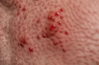
Scar and tattoo seepage
Invasion of scars might take place. Scars from previous trauma, surgery, venipuncture, or tattoo might become penetrated and show a red or purple color. These sores might hurt.
The advancement of sarcoidal sores within tattoos has actually been well recorded, although hypersensitivity granulomatous reactions to tattoo pigment need to thought about in the differential diagnosis and should be thought about in the context of the general scientific discussion of the sarcoid patient. Certain tattoo pigment colors might be targeted more than others, even within the exact same patient with a multicolored tattoo.