What is Vitreomacular Traction Syndrome?
Vitreomacular traction (VMT) syndrome is an eye condition involving the vitreous, the clear gel that fills the within the eyeball. It is a vision-threatening condition and can cause symptoms varying from lowered and blurred vision to distorted and blacked-out main vision. A lot of patients who have VMT syndrome experience some difficulty doing day-to-day tasks that require sharp vision, such as or playing computer game.
Vitreomacular traction can cause a macular hole. A macular hole will misshape and blur the vision and, in most cases, need to be resolved surgically in order to minimize the likelihood of long-term vision loss.
To comprehend the mechanical reasons for VMT, we must first familiarize ourselves with posterior vitreous detachment is and how it connects to VMT.
Posterior Vitreous Detachment (PVD)
The vitreous is a clear, gel-like substance (think of a jellyfish) that fills about 80 percent of the inside of the eyeball. The vitreous helps the eye maintain its round shape. There are millions of fine fibers linked within the vitreous that are connected to the surface of the retina– the eye’s light-sensitive tissue lining the back, inner wall of the eyeball. As the vitreous shrinks (usually due to aging) it ends up being more liquefied, forming pockets of fluid within the vitreous which results in a contraction (syneresis) of the vitreous. Typically, not long after this procedure starts, the vitreous separates from the retina entirely– this is called Posterior Vitreous Detachment– a typical condition that typically impacts individuals over age 50. Once the vitreous has actually separated, the condition is virtually totally safe.
What Causes Vitreomacular Traction?
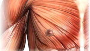
Vitreomacular traction occurs in cases where, rather of totally detaching, the vitreous adheres (vitreomacular adhesion) to the main vision part of the retina– the macula. As only part of the vitreous efforts to pull away, a tugging or pulling happens in the area of adhesion, causing actual physical modifications and distortion of the macula itself. (see diagram).
This relentless pulling on the macula typically causes swelling in its layers, a condition called Cystoid Macular Edema (CME). CME can cause blurring and often distortion of the vision.
How common is Vitreomacular Traction Syndrome?
VMT just takes place in about 1 in 4400 individuals. The incident of VMT in patients with diabetic retinopathy, age-related macular degeneration, and other macular diseases is much higher. It occurs in women slightly regularly than males and can happen at any age, in any race.
How is Vitreomacular Traction diagnosed?
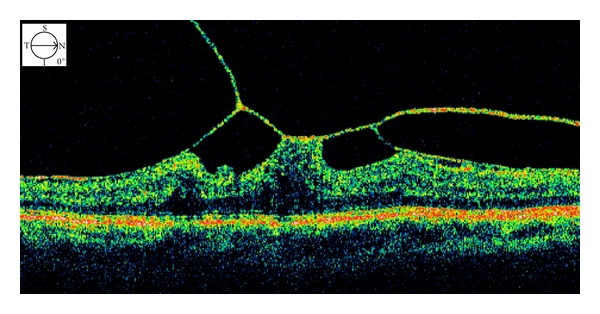
Optical Coherence Tomography (OCT) is a retina/macula scanner that provides an automated, segmented representation of the vitreous and macular layers. This technology has been used in vitreo-retina care for years. This non-invasive diagnostic research can show the retina professional a cross-section view of the vitreous and macula, demonstrating the quantity of involvement and stress on the macula brought on by VMT. OCT has actually become an exceptionally vital tool in identifying and managing macular issues like VMT.
Fluorescein angiography is a valuable test that supplies information about the condition of the retina. This dye study provides the eye physician a “real-time” look at the blood circulation in the retina. It is also useful in discovering any swelling, like CME, that may be present in the macula due to VMT.
Treatment for Vitreomacular Traction Syndrome
Observation
Prior to the development of Optical Coherence Tomography, it was believed that only 11% of eyes with VMT were naturally dealt with by spontaneous detachment of the vitreous. Now that we have better diagnostic tools, it’s most likely more detailed to 20% of VMT cases spontaneously solve.
“Wait and see” is an alternative in some cases, nevertheless, symptoms need to be closely monitored with day-to-day Amsler grid tests and follow-up sees to the ophthalmologist.
Vitreomacular Traction Syndrome and Surgery
If VMT is threatening a macular hole, a pars plana vitrectomy is typically advised. This is a typical retina surgery involving removal of the vitreous. The retina professional surgeon inserts a tiny, needle-like instrument, called a vitrector, into the eye. This instrument will aspirate (suck out) the vitreous fluid. The specialist will then fill up the eye with a special saline solution that carefully resembles the natural vitreous fluid in the eye.
The surgical elimination of the vitreous alleviates the traction on the macula and with time it will go back to its normal structure and shape.
Ocriplasmin
Jetrea is an intravitreal eye-injected drug to deal with Vitreomacular Adhesions. Jetrea is the very first and only FDA-approved medication for the treatment of VMA and its offers a much needed alternative to vitrectomy. The drug works by dissolving the protein fibers that create the adhesion between the vitreous and the macula. Dissolving this connection safely completes the detachment of the vitreous from the macula and eases the traction.