An ultrasound can be interesting for the parents! An ultrasound is a picture that is formed by utilizing sound waves. In order to develop the image, the individual doing the scan spreads a conductive gel on the mother’s belly, then moves a small handheld scanner over the gel. The gel permits sound waves to be passed through the uterus, and after that they recuperate, producing an image. The ultrasound doesn’t harm the mom or the baby, and can provide an exciting peek into the womb. A vaginal ultrasound might also be done, based on the number of weeks pregnant you are. For those who are getting an early ultrasound, you might wonder what happens at 8 week ultrasound and how your baby is developing at this stage.
Why May You Need to Do an Ultrasound at 8 Weeks?
This ultrasound is frequently a terrific moment for parents. It is the most common time for women to have the first ultrasound, simply to make sure that everything is fine. Some ultrasounds might be carried out for other factors; here are a few of them:
- An ultrasound can eliminate an ectopic pregnancy or other problems.
- An ultrasound can verify the presence of multiple pregnancies.
- This ultrasound can inspect the size of the embryo, and validate that there is a heart beat.
- A “dating scan” can access gestational age.
- An ultrasound can determine the health of the ovaries and fallopian tubes.
- If a mom is bleeding, the ultrasound can assist determine the cause.
What Happens at the 8 Week Ultrasound?
How it is done:
Health Support: This Vitamin K2 + D3 Complex is essential for bone density, cardiovascular health, and immune function. It’s a highly-rated formula for those looking to maintain optimal nutrient levels. You can find it on Amazon.
In some cases the ultrasound is done with the wand throughout your belly, as described above. However that might make things harder to see. In that case, a vaginal probe can be used to conduct the ultrasound. This is called a transvaginal ultrasound. It is carried out with a small wand that is placed in the vagina and pushed versus the cervix in order to get a photo from that instructions. Simply as with the other method, the ultrasound is not dangerous and does not hurt, though you may feel some pressure. The majority of women find that the pressure is barely noticeable, specifically when they get to see their baby on the screen.
What you can see:
This first ultrasound gives you a great deal of details, consisting of how the umbilical cord is working, the size of the placenta, the size of the embryoand the heart rate. You may likewise have the ability to tell if you have multiple babies therein. The ultrasound can confirm that everything is healthy and progressing as it must be.
After ultrasound on 8th week of pregnancy, others may be scheduled to make sure that the baby is growing as it must be. Some women choose the transvaginal ultrasound since they are not required to have a full bladder for the scan to work. With the scan over the belly, the doctor may ask a lady to have a complete bladder in order to “raise” the uterus up a bit. Luckily, the later the pregnancy advances, the less likely the doctor will be to want you to have a full bladder at the time of the ultrasound.
Health Support: This high-absorption Magnesium Glycinate (200 mg) is gentle on the stomach and supports muscle relaxation, better sleep, and metabolic health. You can find this trusted formula on Amazon.
The following video shows how your baby appears like at 8 week ultrasound:
How Is Your Baby Developing at Week 8
At this point whatever that an adult human has is now present in the embryo. This implies that it is not an embryo and can be called a fetus. A strong fetal heartbeat must be clear on the ultrasound, and the heart rate must reach between 140 and 170 beats per minute by the 9th week of pregnancy.
Those who do not have a strong heart beat will need to have a follow-up ultrasound to validate the medical diagnosis due to the fact that the fetus may not be practical. In this case, you will have two choices: to miscarry naturally when the time comes, or to undergo a procedure called dilation and curettage, or D&C. About half of women who go through this kind of early pregnancy loss decide to miscarry naturally, while the other half go with the D&C.
At this moment, the baby currently has tiny arms and legs, in addition to buds where the hands and feet will be. It’s about 18 mm in length, the face is beginning to take shape, and the internal organs are forming. The heart is already beating. There is even a small mouth, nostrils, tooth buds, and the beginning of eyes. The baby is moving quite a bit currently, though mama can’t feel it yet.
Image of 8 Week Ultrasound
It is the first good look you will get of your baby! Here’s what to anticipate.
Health Support: The WHOOP 5.0 Activity Tracker offers a comprehensive, 24/7 analysis of your body’s recovery, sleep patterns, daily strain, and key health biometrics. It’s an invaluable tool for anyone looking to optimize their energy, manage stress, and understand what their body truly needs. You can find it on Amazon.
Image 1: As you can see from this photo, the baby’s body is in fact starting to appear like a human. You can see little legs and arms, the head (which is much bigger than the body) and the space where your baby is drifting around.
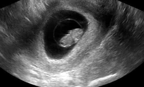
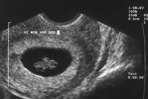
Image 2: This is a more close up picture of the baby form a different angle. This view reveals a much closer representation of the little body, consisting of that round head and belly.
More Notes on Your hCG Level at 8 Week Pregnancy
In a normal pregnancy, hCG levels are a marker of how things are advancing. A good hCG level will peak at 8 to 12 weeks of pregnancy, then gradually decline till it reaches a lower level. When a woman had bloodwork done and the results of the hCG test are doubtful, an ultrasound can help the doctor decide if the pregnancy is viable or not. After five to six weeks of pregnancy, an ultrasound is thought about more accurate than hCG levels in determining how viable the pregnancy really is.