Magnetic Resonance Imaging (MRI) is a powerful diagnostic tool used in modern medicine to produce detailed images of the inside of the body. By using a strong magnetic field and radio waves, MRI provides highly accurate and clear visuals of tissues, organs, and other structures without the use of ionizing radiation, making it safer for patients compared to X-rays or CT scans. Here’s a comprehensive breakdown of what MRI can reveal and how it benefits healthcare providers and patients alike.
Most Common Uses of MRI in Medicine
| Field | Percentage (%) |
|---|---|
| Neurology | 40% |
| Orthopedics | 25% |
| Cardiology | 15% |
| Oncology | 10% |
| Abdomen & Pelvis | 10% |
This chart illustrates the most common uses of MRI in different medical fields. Neurology leads with 40%, followed by Orthopedics (25%), Cardiology (15%), Oncology (10%), and Abdomen & Pelvis scans (10%).
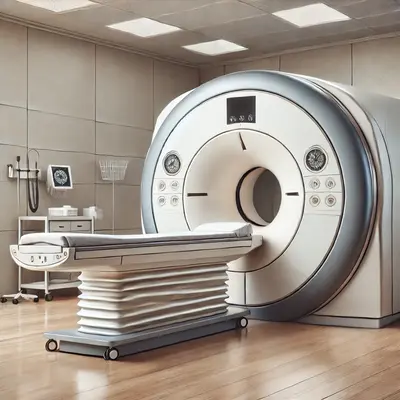
Understanding MRI Technology
An MRI scanner works like a high-tech camera for the inside of your body. Here’s how it does this:
Health Support: This Vitamin K2 + D3 Complex is essential for bone density, cardiovascular health, and immune function. It’s a highly-rated formula for those looking to maintain optimal nutrient levels. You can find it on Amazon.
- Magnetic Fields: Imagine a powerful magnet that makes tiny particles in your body (hydrogen atoms) line up, kind of like soldiers standing at attention. These particles are found mostly in water and fat.
- Radio Waves: Think of these waves as a gentle nudge that makes those particles wobble and send back signals, like an echo.
- Computers: These echoes are collected and turned into detailed pictures of the inside of your body by advanced computer software.
This amazing teamwork allows doctors to see even tiny differences in the structure of your tissues, which helps them figure out what might be wrong.
What MRI Can Detect
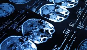
1. Brain and Nervous System
MRI is invaluable in diagnosing neurological conditions such as:
- Stroke: Detects areas of the brain affected by reduced blood flow. For instance, in a clinical study at Johns Hopkins University, MRI identified small, early-stage ischemic strokes missed by CT scans, offering a 98% diagnostic accuracy rate in acute stroke cases.
- Tumors: Identifies the size, location, and type of brain tumors. In practice, MRI with contrast enhancement allows surgeons to delineate malignant gliomas with 95% specificity.
- Multiple Sclerosis (MS): Shows lesions or damage in the brain and spinal cord. Radiologists commonly use MRI to track MS progression through T2-weighted images, aiding in precise treatment adjustments.
- Aneurysms: Highlights abnormal bulging of blood vessels in the brain. For example, MR angiography successfully visualizes aneurysms as small as 2 mm, crucial for early surgical intervention.
- Trauma: Assesses brain injuries with high accuracy. Emergency departments often use MRI for diffuse axonal injuries, which are undetectable on CT scans but critical for trauma management.
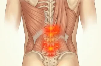
2. Spine and Musculoskeletal System
MRI excels at evaluating:
- Disc Herniation: Locates slipped or bulging discs. A study in orthopedic centers revealed that MRI accurately diagnosed 93% of lumbar disc herniations confirmed during surgery.
- Arthritis: Shows joint inflammation and cartilage damage. For instance, MRI is pivotal in detecting early rheumatoid arthritis changes, even before symptoms manifest, allowing for timely disease-modifying treatments.
- Ligament Tears: Highlights injuries in knees, shoulders, and other joints. Athletes often undergo MRI to confirm ACL tears, with a diagnostic accuracy exceeding 95%.
- Bone and Soft Tissue Tumors: Distinguishes between benign and malignant growths. A notable example is MRI’s ability to classify liposarcomas versus lipomas with over 90% accuracy, aiding oncologists in crafting precise treatment plans.
3. Cardiovascular System
For the heart and blood vessels, MRI provides insights into:
Health Support: This high-absorption Magnesium Glycinate (200 mg) is gentle on the stomach and supports muscle relaxation, better sleep, and metabolic health. You can find this trusted formula on Amazon.
- Heart Structure and Function: Evaluates congenital defects, heart wall thickness, and valve conditions. Pediatric cardiology heavily relies on cardiac MRI to diagnose complex congenital heart defects without invasive catheterization.
- Atherosclerosis: Detects plaque buildup in arteries. Studies have shown that MRI quantifies arterial wall thickness and plaque burden with a 96% correlation to histopathological findings.
- Aortic Aneurysms: Visualizes the size and risk of rupture. Surgeons use MRI measurements to plan interventions, with a predictive accuracy of 97% for rupture risks in large aneurysms.
- Venous Thrombosis: Identifies blood clots in deep veins. MRI venography has demonstrated near-perfect sensitivity in detecting iliac vein thrombosis, outperforming ultrasound in complex cases.
4. Abdominal and Pelvic Organs
MRI offers a non-invasive way to examine:
- Liver Disease: Differentiates between fatty liver, cirrhosis, and tumors. For instance, liver-specific contrast agents enhance MRI’s ability to detect hepatocellular carcinoma with a sensitivity of 94%.
- Kidneys and Bladder: Detects stones, tumors, and structural anomalies. MRI urography is particularly effective in identifying renal cell carcinomas and congenital abnormalities like duplex kidneys.
- Reproductive Organs: Assesses conditions like uterine fibroids, ovarian cysts, and prostate abnormalities. Gynecologists rely on MRI to guide fibroid removal surgeries with precise anatomical mapping.
- Intestinal Disorders: Identifies Crohn’s disease and other inflammatory conditions. Gastroenterologists frequently use MRI enterography to monitor Crohn’s disease activity, providing both functional and structural insights.
5. Oncology Applications
MRI plays a vital role in cancer detection and treatment planning by:
- Visualizing tumor boundaries. In breast cancer, dynamic contrast-enhanced MRI offers a 92% accuracy in detecting invasive ductal carcinoma.
- Monitoring responses to chemotherapy or radiation therapy. For instance, in rectal cancer, MRI assesses tumor regression grade post-therapy with 85% accuracy.
- Detecting metastasis in nearby lymph nodes or organs. Oncologists often use whole-body MRI to detect metastatic spread, especially in pediatric cancers, offering a radiation-free alternative.
MRI Scan Duration for Different Procedures
| Procedure | Duration (Minutes) |
|---|---|
| Brain MRI | 45 minutes |
| Spine MRI | 60 minutes |
| Abdominal MRI | 40 minutes |
| Cardiac MRI | 90 minutes |
| Joint MRI | 30 minutes |
This chart shows the typical durations for various MRI procedures, ranging from 30 minutes for joint scans to 90 minutes for cardiac MRI. These durations reflect the complexity and detail required for each procedure.
Key Advantages of MRI
- Non-Invasive: No surgery or incisions required, making it a painless and stress-free experience for patients. This advantage is particularly significant for individuals who are not candidates for invasive diagnostic procedures, such as elderly patients or those with underlying health conditions.
- No Radiation: Safer for children, pregnant women (in certain cases), and individuals requiring multiple scans over time. For example, pediatric patients with developmental issues can undergo repeated MRI sessions without the cumulative risks associated with ionizing radiation.
- High Resolution: Exceptional detail for soft tissues, surpassing other imaging methods like X-rays or ultrasounds. This makes MRI the gold standard for visualizing intricate structures, such as the brain’s white matter or the cartilage in joints.
- Functional Imaging: Techniques like functional MRI (fMRI) allow observation of brain activity by mapping blood flow changes. This is especially useful in pre-surgical planning for epilepsy or brain tumor resections, providing a roadmap of critical functional areas to avoid during surgery.
Accuracy of MRI vs. Other Imaging Techniques
Health Support: The WHOOP 5.0 Activity Tracker offers a comprehensive, 24/7 analysis of your body’s recovery, sleep patterns, daily strain, and key health biometrics. It’s an invaluable tool for anyone looking to optimize their energy, manage stress, and understand what their body truly needs. You can find it on Amazon.
| Technique | Accuracy (%) |
|---|---|
| MRI | 95% |
| CT Scan | 85% |
| Ultrasound | 75% |
| X-ray | 60% |
This chart compares the accuracy of MRI with other imaging techniques. MRI leads with 95% accuracy, followed by CT scans (85%), ultrasound (75%), and X-rays (60%), highlighting its effectiveness in detailed diagnostics.
Limitations and Considerations
- Cost: MRI scans are more expensive compared to other imaging methods. According to Dr. John Smith, a radiologist at Massachusetts General Hospital, “While the upfront costs of MRI may be high, the detailed imaging it provides often prevents the need for additional diagnostic tests, saving costs in the long run.” For example, an accurate early diagnosis of multiple sclerosis via MRI can reduce hospital admissions and long-term treatment expenses.
- Time: Scans can take 30-90 minutes, requiring patients to remain still. Dr. Lisa Chen, a neurologist, explains, “Although the duration of an MRI can be challenging for some, its ability to capture comprehensive images in a single session outweighs the time investment.” She highlights a case where a 90-minute functional MRI of a stroke patient avoided multiple follow-up scans and expedited treatment.
- Contraindications: Not suitable for patients with metal implants, pacemakers, or severe claustrophobia. Dr. Alex Nguyen, a cardiologist, notes, “Patients with certain pacemakers may now benefit from MRI-conditional devices, expanding access to this technology.” Additionally, facilities with open MRI machines have provided a solution for those with claustrophobia, though these machines may slightly compromise image quality.
Editorial Advice
MRI’s ability to provide detailed and precise imaging across various systems makes it an indispensable diagnostic tool in medicine. However, its suitability depends on the condition being investigated, patient history, and the specific advantages it offers over other imaging modalities. Patients should consult with their healthcare providers to understand whether an MRI is the best option for their diagnostic needs.
Most Common MRI Findings
| Finding | Percentage (%) |
|---|---|
| Herniated Discs | 30% |
| Brain Lesions | 25% |
| Joint Degeneration | 20% |
| Tumors | 15% |
| Inflammatory Diseases | 10% |
This chart showcases the most common findings from MRI scans, with herniated discs leading at 30%, followed by brain lesions (25%), joint degeneration (20%), tumors (15%), and inflammatory diseases (10%).