This short article plans to manifest parts of the eye and their functions. Understanding the different parts of the eye can help you comprehend how you see and what you can do to help keep the eye functioning correctly.
The eye is one of the most intricate parts of the body. The different parts of the eye permit the body to take in light and perceive objects around us in the proper color, information and depth. This allows people to make more educated decisions about their environment. If a part of the eye becomes damaged, you might not be able to see efficiently, or lose your vision completely, states iytmed.com. What are the parts of the eye? Which part is not work properly when we suffer various vision problems like myopia and glaucoma? Which part produces tears?
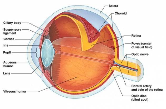
Parts of the Eye and Their Functions
There are numerous physical and chemical aspects that make up the eye. The eye is likewise greatly included with the nerve system, which permits the brain to take in information from the eyes and make the proper choices on how to act on this info. The nerves need to be kept in prime condition or the brain might begin to get false images, or you will not take in enough information to get a precise perception of your environment.
Health Support: This Vitamin K2 + D3 Complex is essential for bone density, cardiovascular health, and immune function. It’s a highly-rated formula for those looking to maintain optimal nutrient levels. You can find it on Amazon.
| Eye Parts | Description and Functions |
|---|---|
| Cornea | The cornea is the external covering of the eye. This dome-shaped layer safeguards your eye from elements that might cause damage to the inner parts of the eye. There are numerous layers of the cornea, developing a difficult layer that provides extra protection. These layers regrow extremely rapidly, helping the eye to eliminate damage more easily. The cornea likewise allows the eye to properly focus on light better. Those who are having trouble focusing their eyes properly can have their corneas surgically improved to remove this problem. |
| Sclera | The sclera is frequently described as the “whites” of the eye. This is a smooth, white layer on the outdoors, but the within is brown and consists of grooves that help the tendons of the eye connect effectively. The sclera provides structure and security for the inner operations of the eye, however is likewise flexible so that the eye can move to seek out things as essential. |
| Pupil | The pupil appears as a black dot in the middle of the eye. This black area is actually a hole that takes in light so the eye can focus on the items in front of it. |
| Iris | The iris is the area of the eye that contains the pigment which gives the eye its color. This area surrounds the pupil, and uses the dilator pupillae muscles to broaden or close the student. This permits the eye to take in basically light depending on how brilliant it is around you. If it is too intense, the iris will diminish the pupil so that they eye can focus better. |
| Conjunctiva Glands | These are layers of mucus which help keep the beyond the eye moist. If the eye dries out it can end up being itchy and painful. It can also become more vulnerable to damage or infection. If the conjunctiva glands become infected the patient will cultivate “pink eye.” |
| Lacrimal Glands | These glands lie on the outer corner of each eye. They produce tears which help moisten the eye when it becomes dry, and flush out particles which aggravate the eye. As tears flush out potentially dangerous irritants, it becomes simpler to focus correctly. |
| Lens | The lens sits directly behind the student. This is a clear layer that focuses the light the pupil takes in. It is held in place by the ciliary muscles, which allow the lens to change shape depending on the quantity of light that strikes it so it can be correctly focused. |
| Retina | The light focuses by the lens will be transferred onto the retina. This is made from rods and cones set up in layers, which will transmit light into chemicals and electrical pulses. The retina lies in the back of the eye, and is linked to the optic nerves that will send the images the eye sees to the brain so they can be analyzed. The back of the retina, known as the macula, will help interpret the details of the item the eye is working to translate. The center of the macula, referred to as the fova will increase the information of these images to a perceivable point. |
| Ciliary Body | Ciliary body is a ring-shaped tissue which holds and controls the motion of the eye lens, and hence, it helps to control the shape of the lens. |
| Choroid | The choroid lies between the retina and the sclera, which provides blood supply to the eye. Similar to any other portion of the body, the blood supply gives nutrition to the various parts of the eye. |
| Vitreous Humor | The vitreous humor is the gel located in the back of the eye which helps it hold its shape. This gel takes in nutrients from the ciliary body, aqueous humor and the retinal vessels so the eye can remain healthy. When debris finds its method into the vitreous humor, it causes the eye to view “floaters,” or spots that cross the vision area that can not be attributed to items in the environment. |
| Aqueous Humor | The aqueous humor is a watery substance that fills the eye. It is divided into two chambers. The anterior chamber is located in front of the iris, and the posterior chamber is straight behind it. These layers allow the eye to preserve its shape. This liquid is drained through the Schlemm canal so that any accumulation in the eye can be removed. If the patient’s aqueous humor is not draining effectively, they can establish glaucoma. |