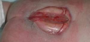
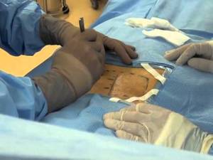
Surgery for spina bifida includes a variety of neurosurgical, orthopedic, and urologic treatments.
Surgical procedures consist of the following:
- Closure of the flaw over the spinal cord
- Back defect reconstruction
- Lower-extremity defect correction
Without closure of the flaw, survival is threatened. Closure may end up being more frequently a prenatal procedure.
Some have actually viewed the connection between spina bifida and hydrocephalus in a unified sense, such that spina bifida is a penalty of fetal hydrocephalus.
Beyond closure, other needed neurosurgical treatments might consist of shunting for hydrocephalus, including regular pressure hydrocephalus. Numerous urologic procedures might be required for the neurogenic bladder.
In addition to neurologic flaws, back defect prevails in patients with spina bifida and is really difficult to filter. Nevertheless, correction and fusion are essential to detain development and problems and are requirements for effective treatment of lower-extremity defects and for decrease of impedances to walking and sitting. The literature has cannot document a substantial quality-of-life enhancement with fusion.
Infection prevails, especially in spina bifida patients with a neurogenic bladder. An enhanced danger exists with any operative treatment. Retethering of the spinal cord frequently happens and might manifest urologically and orthopedically. Any presumed weather change in muscle status, which have to be kept an eye on serially, or urologic status might signify retethering.
Back defect reconstruction might be particularly challenging since of posterior element deficiencies. These can cause instrumentation and blend failures, infection from the neurogenic bladder, and distal ulcers from insensate skin. Anterior procedures are being integrated more regularly with the posterior technique to produce an adequate combination.
Lower-extremity procedures are necessitated by muscle imbalance forces. These treatments, particularly hip reduction, have actually been the most questionable. Opinions vary as to whether they aid in seating and ambulation. Gait analysis has been utilized to examine particular prospects who have motor strength in the quadriceps in the variety of “excellent plus” and who likewise have hip dysplasia or dislocation.
Treatments distal to the hip typically resemble those for polio however generally are focused on keeping the capability to brace the limb to make the most of ease of access and independence. These treatments are directed toward decreasing deformity, launching contractures, and balancing muscle forces from the variable neurologic sores.
The orthopedic cosmetic surgeon has a large function in the treatment of patients with spina bifida, in addition to medical intervention. This includes long-term monitoring of neurologic status, motor stamina, and joint range of movement. Since many patients require some degree of bracing, examination for skin inflammation and breakdown and observation of ambulation to assess the practical utility of the braces are practical in finding any change in condition or the need for treatment modification.
Spina Bifida Surgery
Surgery is the most common treatment for spina bifida and its complications. Most children with severe spina bifida need a series of operations.
- The first, which typically is done in the first 48 hours of the child’s life, includes tucking the exposed spine and nerve roots back into the surrounding membrane, closing the problems in cord and membrane, and covering the injury with muscle and skin flaps drawn from either side of the back.
- Succeeding spina bifida surgeries involve correction of defects. This may include cutting tendons or ligaments to launch contractures and/or rebalancing muscles around the included joint. When regular assessments indicate that the individual’s performance is declining while a physical defect becomes worse, surgery must be thought about.
Urologic surgery is typically essential, due to the fact that unresisted contracture restricts the capability of the bladder to hold sufficient urine to space out emptying and may obstruct circulation from the kidneys. Neglected, this can result in kidney failure, which can cause premature death.
In the 1990s, pioneering specialists developed a technique for repairing the spine while the fetus is still in the womb. The reasoning behind this is that the longer the spine is exposed to outdoors aspects, even in the womb, the greater the possibility for damage to the cord and nerve roots. Hence, earlier surgery might prevent a few of the damage that has currently occurred by the time the baby is born.
- Initial outcomes have actually been excellent in that infants who went through prenatal surgery are less likely than babies who went through surgery at birth to require a shunt for drain of hydrocephalus fluid.
This surgery is not without danger, obviously; like any operation, it carries considerable dangers. This surgery also dramatically increases the threat of premature birth, which entails its own set of risks for the baby. - It is too early to tell yet whether this operation deserves the risks it involves. Scientists are observing the children who have actually undergone this surgery as they grow, to see whether they do much better than children who go through the traditional spina bifida surgery.
Hydrocephalus normally is filtered by positioning of a shunt. A shunt is a special tube surgically put in the head and under the skin down into the chest or abdomen. The shunt drains excess fluid from the brain into the abdomen, where it can be eliminated without harm.


