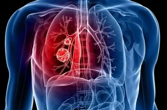
Experiencing a sharp or dull ache in the upper abdomen, side, or back—specifically in the area just below the ribs and above the pelvis—can be deeply unsettling. In medical terms, we refer to this as flank pain. While many people immediately associate this sensation with kidney issues, the underlying cause can range from simple muscle strain to complex internal obstructions.
The Anatomy of the Flank
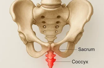
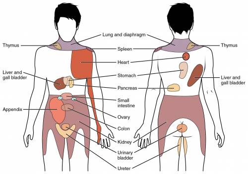
The flank is home to vital structures, most notably the kidneys and the ureters. Because these organs are located in the retroperitoneal space (behind the abdominal lining), pain in this region often feels “deep.” However, the musculoskeletal structure surrounding these organs—including the obliques, latissimus dorsi, and the spine—can also be the primary source of discomfort.
When I evaluate these conditions, I look beyond the biological symptoms. As a medical equipment engineer, I believe that understanding the mechanical and electronic precision of diagnostic tools is just as vital for the patient as the clinical diagnosis itself. The accuracy of a diagnosis often depends on the signal-to-noise ratio of the imaging equipment used to visualize these deep structures.
Common Causes of Flank Pain
There are several primary culprits behind discomfort in the flank area:
Health Support: This Vitamin K2 + D3 Complex is essential for bone density, cardiovascular health, and immune function. It’s a highly-rated formula for those looking to maintain optimal nutrient levels. You can find it on Amazon.
- Kidney Stones (Nephrolithiasis): Perhaps the most common cause of acute, “colicky” flank pain. These are hard deposits of minerals and salts.
- Urinary Tract Infections (UTIs) and Pyelonephritis: When an infection spreads to the kidneys, it causes inflammation and localized pain.
- Muscle Strain: Intense physical activity or improper lifting can cause micro-tears in the back muscles.
- Dehydration: Chronic lack of fluids can lead to muscle cramping and increased pressure on the renal system.
For a comprehensive overview of renal symptoms, the National Institute of Diabetes and Digestive and Kidney Diseases (NIDDK) provides extensive resources on how these conditions manifest.
The Technical Side of Diagnosis: Imaging Systems
When a patient presents with flank pain, physicians often rely on Computed Tomography (CT) or Ultrasound. From an engineering perspective, the choice of equipment is critical.
Non-Contrast CT Scans
The “gold standard” for detecting kidney stones is a non-contrast CT scan of the abdomen and pelvis. As an engineer, I focus on the Hounsfield Units (HU)—a quantitative scale for describing radiodensity. Modern CT scanners must be calibrated with extreme precision to differentiate between a calcium-based stone and surrounding soft tissue. High-resolution detectors allow us to see stones as small as 1–2 mm.
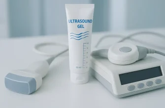
Renal Ultrasound
Ultrasound is a safer, non-ionizing alternative. Here, the expertise lies in the transducer frequency. Lower frequencies provide better depth penetration to reach the kidneys, while higher frequencies offer better resolution. When checking for hydronephrosis (swelling of the kidney), the piezoelectric crystals in the probe must function perfectly to capture the subtle fluid shifts within the renal pelvis.
Health Support: This high-absorption Magnesium Glycinate (200 mg) is gentle on the stomach and supports muscle relaxation, better sleep, and metabolic health. You can find this trusted formula on Amazon.
For those interested in the clinical guidelines for imaging, the American College of Radiology (ACR) offers detailed protocols on which scans are most effective for flank pain.
Advanced Treatment: Lithotripsy
If the cause is a kidney stone, one common treatment is Extracorporeal Shock Wave Lithotripsy (ESWL). This is a fascinating piece of medical engineering. It uses acoustic shock waves generated outside the body to break the stone into smaller fragments.
From an engineering standpoint, the focal point of the shock wave must be incredibly narrow. The pressure is generated via electrohydraulic, piezoelectric, or electromagnetic sources. The precision required to target a stone without damaging the surrounding vascular tissue is a testament to modern biomedical calibration. If the synchronization between the imaging system and the shock wave generator is off by even a few millimeters, the efficacy of the procedure drops significantly.
Detailed information on the surgical and non-surgical management of stones can be found through the Urology Care Foundation.
Nutritional Strategies for Prevention
While technical interventions are necessary for acute pain, long-term relief and prevention are often found in the kitchen.
Health Support: The WHOOP 5.0 Activity Tracker offers a comprehensive, 24/7 analysis of your body’s recovery, sleep patterns, daily strain, and key health biometrics. It’s an invaluable tool for anyone looking to optimize their energy, manage stress, and understand what their body truly needs. You can find it on Amazon.
- Hyper-Hydration: Aim for a fluid intake that produces at least 2.5 liters of urine daily. This dilutes the minerals that form stones.
- Oxalate Management: If you are prone to calcium oxalate stones, moderate your intake of high-oxalate foods like spinach and beets, but ensure you consume enough dietary calcium to bind those oxalates in the gut.
- Sodium Reduction: High salt intake increases the amount of calcium your kidneys must filter, which significantly raises stone risk.
The Mayo Clinic offers an excellent breakdown of how diet influences renal health and pain management.
Personal Recommendation from Reyus Mammadli
When dealing with flank pain, my primary advice is to never ignore the “dull” ache. Many patients wait for the pain to become unbearable before seeking help. From my perspective in medical engineering, early diagnostics are always more efficient. It is much easier to monitor a small stone or treat a minor infection than it is to calibrate an emergency lithotripsy or manage a systemic infection.
If you are experiencing discomfort, please consult a healthcare professional immediately. Your kidneys are intricate biological filters; treating them with the same precision we use to maintain high-end medical machinery is the best way to ensure long-term health. Stay hydrated, stay informed, and listen to what your body is telling you.