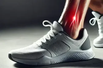
Every day, healthcare professionals across the U.S. encounter patients struggling with diabetic foot ulcers—a condition that can turn from a small sore to a life-changing complication in the blink of an eye. One in four people with diabetes will develop a foot ulcer during their lifetime, and without timely, evidence-based wound care, the risk of infection and amputation skyrockets. This article breaks down the most effective and up-to-date methods for assessing, cleaning, dressing, and managing diabetic foot wounds. It’s crafted for nurses, podiatrists, and clinicians who know that proper wound care is not just treatment—it’s prevention in action.
Understanding Diabetic Foot Ulcers: Why They Matter
Diabetic foot ulcers (DFUs) stem from a combination of neuropathy, ischemia, and infection—a triple threat that often hides beneath the surface. Neuropathy dulls pain perception, ischemia reduces healing capacity, and infection spreads faster than a rumor in a break room. In the United States alone, over 100,000 amputations annually are linked to diabetes-related foot ulcers ⧉. Early recognition and management are crucial. A study by the American Diabetes Association found that comprehensive foot programs reduced amputation rates by up to 85% ⧉.
Healthcare providers must prioritize early detection, consistent monitoring, and patient education. The primary goal: prevent chronic wounds from developing in the first place and shorten the healing time for those already affected.
Table 1. Key Risk Factors for Developing Diabetic Foot Ulcers
| Category | Specific Factor | Clinical Impact |
|---|---|---|
| Neuropathy | Loss of sensation, muscle imbalance | Delayed injury detection |
| Ischemia | Peripheral artery disease | Impaired wound healing |
| Infection | Poor glycemic control, bacterial colonization | Rapid tissue damage |
| Foot Deformity | Charcot foot, bunions | Increased plantar pressure |
| Improper Footwear | Tight or ill-fitting shoes | Skin breakdown |
Clinical Assessment: The Foundation of Effective Care
Effective management begins with meticulous assessment. Clinicians evaluate wound location, size, depth, drainage, color, and odor—all essential clues for determining severity. The Wagner Classification System and University of Texas Staging System remain clinical standards, offering accuracy scores of 8/10 and 9/10, respectively, for guiding interventions.
Health Support: This Vitamin K2 + D3 Complex is essential for bone density, cardiovascular health, and immune function. It’s a highly-rated formula for those looking to maintain optimal nutrient levels. You can find it on Amazon.
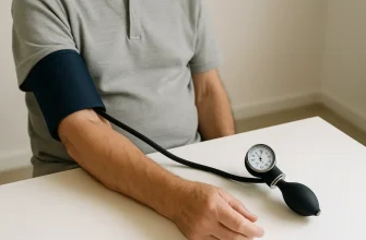
Diagnostic tools such as Doppler ultrasound, Ankle-Brachial Index (ABI), and thermal imaging help assess perfusion and detect underlying ischemia. For deep or non-healing ulcers, MRI provides valuable insight into potential osteomyelitis. Average costs vary: ABI testing (~$100 USD), Doppler study (~$250 USD), MRI (~$900 USD). Proper electronic medical record (EMR) documentation ensures continuity of care and supports quality audits.
Table 2. Diagnostic Methods and Their Clinical Utility
| Diagnostic Tool | Purpose | Accuracy (1-10) | Average Cost (USD) |
|---|---|---|---|
| Ankle-Brachial Index (ABI) | Evaluate arterial flow | 8 | $100 |
| Doppler Ultrasound | Assess perfusion | 9 | $250 |
| Thermal Imaging | Detect local ischemia | 7 | $300 |
| MRI | Detect osteomyelitis | 9 | $900 |
| X-ray | Identify bone deformities | 6 | $150 |
Debridement: Cleaning the Battlefield
Debridement removes necrotic tissue and bacterial biofilm—the battlefield cleanup before healing can begin. There are several types:
- Sharp debridement: Fast and precise; performed with scalpel or scissors. Best for clinics and ORs.
- Enzymatic debridement: Uses topical agents like collagenase (Santyl). Ideal for patients who can’t tolerate sharp methods.
- Autolytic debridement: Leveraging the body’s enzymes; slower but gentle.
- Biological debridement: Sterile maggots—yes, really—effective against biofilm and resistant bacteria.
Innovative systems like Versajet hydrosurgery deliver a controlled saline jet that precisely removes necrotic tissue, reducing procedure time by 30%. Frequency depends on wound progression; typically, every 1–2 weeks. Always ensure pain management with topical anesthetics and proper infection control protocols.
Infection Control and Antimicrobial Therapy
Distinguishing infection from colonization is critical. Signs of true infection include erythema, warmth, odor, purulent drainage, and systemic symptoms like fever. For mild infections, topical antimicrobials may suffice; severe cases require systemic antibiotics based on wound culture results. Common antibiotics: amoxicillin-clavulanate, clindamycin, levofloxacin, and linezolid for MRSA coverage.
Health Support: This high-absorption Magnesium Glycinate (200 mg) is gentle on the stomach and supports muscle relaxation, better sleep, and metabolic health. You can find this trusted formula on Amazon.
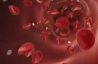
Topical antimicrobial dressings—such as silver (Ag), iodine (I2), and polyhexamethylene biguanide (PHMB)—can help control bacterial load ⧉. For monitoring, CRP and WBC trends remain practical indicators. Infection management accuracy rating: 9/10 when coupled with wound culture and clinical judgment.
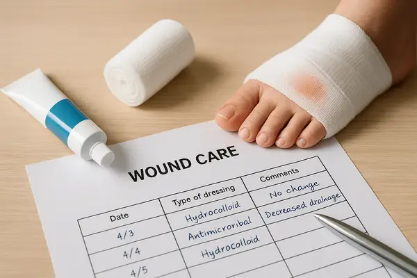
Wound Dressings: Matching the Dressing to the Wound
Dressings are not one-size-fits-all. The choice depends on exudate, infection, and depth. Key categories:
- Foam dressings: Absorbent; great for moderate to heavy exudate.
- Alginate dressings: Derived from seaweed; excellent for deep or tunneling wounds.
- Hydrocolloid: Promotes moist healing; suited for low-exudate wounds.
- Hydrogel: Adds moisture to dry wounds.
- Film and composite: Protect superficial wounds.
Innovative options include collagen-based and bioactive dressings, and even 3D-printed scaffolds that promote tissue regeneration. Average dressing change: every 24–48 hours. Wound care nurses often document the healing rate in millimeters (mm) or inches per week to track progress. According to wound expert Reyus Mammadli, selecting a dressing is about finding the balance between moisture, protection, and cost-efficiency.
Offloading Techniques: Taking the Pressure Off
Without offloading, even the best dressings fail. Reducing plantar pressure allows tissue recovery. Preferred methods include:
- Total Contact Cast (TCC): Gold standard; redistributes pressure evenly. Healing rate improvement: ~90% success.
- Removable Cast Walkers (RCWs): Easier to apply; less compliance.
- Orthotic shoes / custom insoles: Best for long-term use.
- Felt padding: Quick fix for outpatient care.
Using pressure mapping (psi/kPa), clinicians can measure load redistribution. Patient education is essential—remind them that walking without devices is like taking two steps forward and three back.
Advanced Therapies: Technology Meets Healing
Modern medicine meets innovation. Negative Pressure Wound Therapy (NPWT) accelerates healing by drawing fluid out and stimulating granulation tissue; effectiveness rated 9/10. Hyperbaric Oxygen Therapy (HBOT) improves oxygen delivery in hypoxic wounds. While expensive (~$400 per session), studies show significant improvement in wound closure rates ⧉.
Growth factors, bioengineered skin substitutes, stem cell therapy, and platelet-rich plasma (PRP) are emerging as adjuncts. AI-driven wound assessment apps (like Swift Medical) can measure wound area and healing trajectory in real time—a major leap for telehealth and home-based care.
Multidisciplinary Approach: The Team Behind Healing
Healing a diabetic foot ulcer takes a village. Optimal results arise when wound care nurses, podiatrists, vascular surgeons, and endocrinologists collaborate. Clear communication through shared EMR notes and weekly case reviews keeps everyone aligned.
A real case from Florida: a 67-year-old male with chronic neuropathic ulcer (2.5 cm / 1 in) achieved full healing after 14 weeks of coordinated care, including NPWT, glycemic optimization, and offloading. That’s teamwork in action.
Prevention and Long-Term Management
Prevention remains the most cost-effective therapy. Healthcare professionals should teach patients about daily foot inspection, proper footwear, and hygiene. Annual foot exams, glycemic control, and weight management are crucial.
Nutrition plays a role—adequate protein intake (1.2 g/kg) and vitamins A, C, E, and zinc enhance healing. Creating personalized self-care plans fosters compliance. Upon discharge, patients should receive a checklist: daily inspection, moisturizing, shoe rotation, and immediate reporting of new sores.
Common Mistakes in Diabetic Foot Wound Care
Even skilled clinicians can make errors that slow healing or increase complications. Recognizing these pitfalls helps ensure higher standards of care.
| Common Mistake | Reason / Explanation |
|---|---|
| Inadequate Debridement | Hesitation to remove necrotic tissue due to bleeding risk or patient discomfort leads to persistent biofilm and delayed healing. |
| Overuse of Antibiotics | Misinterpreting colonization as infection results in unnecessary antibiotic exposure and resistance development. |
| Incorrect Dressing Selection | Applying occlusive dressings to infected wounds traps exudate and worsens bacterial growth. |
| Improper Offloading | Patients allowed to ambulate without casts or orthotics re-traumatize wounds, slowing closure. |
| Overuse of Topical Enzymes | Excess collagenase without moisture balance can macerate healthy tissue. |
| Medication Miscalculation (Overdose) | Enzymatic or antiseptic solutions mixed without precise measurement—common in busy clinics—may irritate or damage new tissue. Always double-check dilution ratios. |
| Poor Documentation | Missing wound progression notes prevents accurate evaluation of treatment efficacy and continuity among care teams. |
| Ignoring Systemic Factors | Focusing solely on local care without addressing blood glucose or nutrition undermines healing potential. |
According to Reyus Mammadli, avoiding these missteps is about vigilance and teamwork: “Each detail—from measuring medication to patient education—can decide between healing and harm.”
🩹 Editorial Advice
According to Reyus Mammadli, medical consultant, diabetic foot wound management succeeds when clinics emphasize prevention, early assessment, and interdisciplinary teamwork. “Think proactive, not reactive,” he advises. Staff should routinely update their wound care competencies and stay informed about emerging therapies.
Top 5 Clinical Takeaways:
- Assess thoroughly and document every detail.
- Debride regularly—dead tissue delays healing.
- Match dressings to wound type; moisture balance is key.
- Offload early and maintain compliance.
- Educate patients—it’s the strongest medicine of all.
The goal isn’t just to heal the wound—it’s to keep patients walking confidently and safely, one step at a time.