Echinococcosis is a parasitic disease brought on by infection with small tapeworms of the genus Echinocococcus. Echinococcosis is categorized as either cystic echinococcosis or alveolar echinococcosis. In this article we will talk about the two main types of the disease, common symptoms in human and how to treat and/or prevent it.
Cystic echinocccosis (CE), likewise called hydatid disease, is brought on by infection with the larval stage of Echinococcus granulosus, a ~ 2-7 millimeter long tapeworm found in dogs (definitive host) and sheep, livestock, goats, and pigs (intermediate hosts). Although the majority of infections in humans are asymptomatic, CE causes damaging, slowly enlarging cysts in the liver, lungs, and other organs that frequently grow undetected and overlooked for many years.
Alveolar echinococcosis (AE) disease is caused by infection with the larval stage of Echinococcus multilocularis, a ~ 1-4 millimeter long tapeworm discovered in foxes, coyotes, and dogs (conclusive hosts). Little rodents are intermediate hosts for E. multilocularis. Although cases of AE in animals in endemic areas are relatively typical, human cases are rare. AE positions a much greater health hazard to people than CE, causing parasitic tumors that can form in the liver, lungs, brain, and other organs. If left untreated, AE can be deadly.
Symptoms
Cystic echinococcosis
Human infection with E. granulosus results in the development of several hydatid cysts located frequently in the liver and lungs, and less frequently in the bones, kidneys, spleen, muscles, central nerve system and eyes.
Health Support: This Vitamin K2 + D3 Complex is essential for bone density, cardiovascular health, and immune function. It’s a highly-rated formula for those looking to maintain optimal nutrient levels. You can find it on Amazon.
The asymptomatic incubation period of the disease can last many years up until hydatid cysts grow to a level that activates clinical signs, nevertheless approximately half of all patients that get medical treatment for infection do so within a couple of years of their preliminary infection with the parasite.
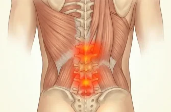
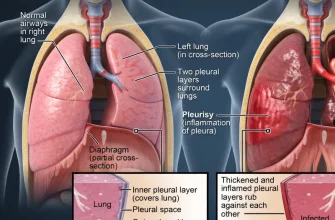
Abdominal pain, nausea and vomiting are typically seen when hydatids occur in the liver. If the lung is impacted, clinical signs include chronic cough, chest pain and shortness of breath. Other signs depend upon the location of the hydatid cysts and the pressure put in on the surrounding tissues. Non-specific signs include anorexia, weight-loss and weak point.
Alveolar echinococcosis
Alveolar echinococcosis is characterized by an asymptomatic incubation period of 5– 15 years and the slow advancement of a main tumour-like sore which is normally located in the liver. Scientific signs consist of weight reduction, abdominal pain, basic malaise and signs of hepatic failure.
Larval metastases may spread either to organs surrounding to the liver (for example, the spleen) or distant areas (such as the lungs, or the brain) following dissemination of the parasite by means of the blood and lymphatic system. If left untreated, alveolar echinococcosis is progressive and deadly.
Health Support: This high-absorption Magnesium Glycinate (200 mg) is gentle on the stomach and supports muscle relaxation, better sleep, and metabolic health. You can find this trusted formula on Amazon.
Echinococcosis Causes and Transmission
A number of herbivorous and omnivorous animals act as intermediate hosts of Echinococcus. They end up being infected by consuming the parasite eggs in contaminated food and water, and the parasite then turns into larval stages in the viscera.
Carnivores act as definitive hosts for the parasite, and host the mature tapeworm in their intestine. They are infected through the consumption of viscera of intermediate hosts that harbour the parasite.
Humans serve as so-called unintentional intermediate hosts in the sense that they get infection in the very same way as other intermediate hosts, but are not associated with sending the infection to the conclusive host.
Numerous distinct genotypes of E. granulosus are acknowledged, some having distinct intermediate host choices. Some genotypes are thought about species unique from E. granulosus. Not all genotypes cause infections in people. The genotype triggering the great majority of cystic echinococcosis infections in people is principally kept in a dog — sheep — dog cycle, yet numerous other domestic animals may likewise be included, consisting of goats, swine, cattle, camels and yaks.
Alveolar echinococcosis generally happens in a wildlife cycle in between foxes, other predators and small mammals (primarily rodents). Domesticated dogs and cats can likewise be infected.
Health Support: The WHOOP 5.0 Activity Tracker offers a comprehensive, 24/7 analysis of your body’s recovery, sleep patterns, daily strain, and key health biometrics. It’s an invaluable tool for anyone looking to optimize their energy, manage stress, and understand what their body truly needs. You can find it on Amazon.
Distribution
Cystic echinococcosis is worldwide distributed and found in every continent other than Antarctica. Alveolar echinococcosis is restricted to the northern hemisphere, in particular to regions of China, the Russian Federation and countries in continental Europe and North America.
In endemic areas, human occurrence rates for cystic echinococcosis can reach more than 50 per 100 000 person-years, and occurrence levels as high as 5% — 10% may happen in parts of Argentina, Peru, East Africa, Central Asia and China. In livestock, the frequency of cystic echinococcosis found in slaughterhouses in hyperendemic areas of South America varies from 20% — 95% of butchered animals.
The greatest frequency is found in backwoods where older animals are slaughtered. Depending on the infected types involved, livestock production losses attributable to cystic echinococcosis arise from liver condemnation and may likewise include decrease in carcass weight, decrease in hide value, decline of milk production, and decreased fertility.
How Is Echinococcosis Diagnosed?
Ultrasonography imaging is the technique of choice for the diagnosis of both cystic echinococcosis and alveolar echinococcosis in human beings. This strategy is generally complemented or confirmed by computed tomography (CT) and/or magnetic resonance imaging (MRI) scans.
Cysts can be by the way found by radiography. Particular antibodies are discovered by various serological tests and can support the medical diagnosis. Biopsies and ultrasound-guided punctures might likewise be performed for differential diagnosis of cysts from tumours and abscesses.
Treatment for Echinococcosis
Both cystic echinococcosis and alveolar echinococcosis are typically expensive and complicated to treat, often needing substantial surgery and/or extended drug therapy. There are 4 choices for the treatment of cystic echinococcosis:
- percutaneous treatment of the hydatid cysts with the PAIR (Puncture, Aspiration, Injection, Re-aspiration) technique;
- surgery
- anti-infective drug treatment
- “watch and wait”.
The option should mainly be based on the ultrasound pictures of the cyst, following a stage-specific technique, as well as on the medical facilities and personnels available.
For alveolar echinococcosis, early diagnosis and radical (tumour-like) surgery followed by anti-infective prophylaxis with albendazole remain the key elements. If the sore is confined, extreme surgery can be curative. Regrettably in many patients the disease is diagnosed at a sophisticated stage. As a result, if palliative surgery is performed without complete and efficient anti-infective treatment, regular regressions will happen.
Early detection of E. granulosus and E. multilocularis infections, especially in low-resource settings, is still needed in addition to the assessment of clinical treatment alternatives. More evaluation and possible commercialization of a vaccine for E. granulosus recombinant oncosphere antigen (EG95) is on trial in sheep to restrain E. granulosus infection of lambs. This could supplement control measures such as the treatment of dogs and culling of older sheep.
Health and Economic Concern
Both cystic echinococcosis and alveolar echinococcosis represent a considerable disease concern. Worldwide, there might remain in excess of 1 million people living with these diseases at any one time. Much of these people will be experiencing severe medical syndromes which are dangerous if left unattended. Even with treatment, people frequently deal with decreased lifestyle.
For cystic echinococcosis, there is approximately 2.2% post-operative death rate for surgical patients and about 6.5% of cases regression after an intervention, thus requiring extended recovery time.
The 2015 WHO Foodborne Disease Burden Epidemiology Reference Group (FERG) approximated echinococcosis to be the cause of 19 300 deaths and around 871 000 disability-adjusted life-years (DALYs) (1) globally each year.
Annual expenses associated with cystic echinococcosis are estimated to be US$ 3 billion for treating cases and losses to the animals industry.
Prevention
Robust monitoring data is basic in order to reveal burden of disease and to evaluate development and success of control programmes. Nevertheless, as for other disregarded diseases which are focused in underserved populations and remote areas, information is specifically limited and will need more attention if control programs are to be executed and measured.
Cystic echinococcosis – hydatid disease
Surveillance for cystic echinococcosis in animals is challenging due to the fact that the infection is asymptomatic in livestock and dogs. Monitoring is also not recognized or focused on by communities or local veterinary services.
Cystic echinococcosis is a preventable disease as it includes domestic animal types as definitive and intermediate hosts. Regular deworming of dogs, enhanced hygiene in the slaughtering of livestock (including the proper damage of infected offal), and public education campaigns have actually been found to lower and, in high-income countries, prevent transmission and minimize the problem of human disease.
Vaccination of sheep with an E. granulosus recombinant antigen (EG95) offers motivating prospects for prevention and control. Small-scale EG95 vaccine trials in sheep show high effectiveness and safety with immunized lambs not becoming infected with E. granulosus.
A programme combining vaccination of lambs, deworming of dogs and culling of older sheep might lead to removal of cystic echinococcosis disease in human beings in less than 10 years.
Alveolar echinococcosis control and prevention
Prevention and control of alveolar echinococcosisis more complex as the cycle involves wild animal types as both definitive and intermediate hosts. Regular deworming of domestic carnivores that have access to wild rodents need to help to minimize the risk of infection in human beings.
Deworming of wild and roaming definitive hosts with anthelminthic baits led to substantial decreases in alveolar echinococcosis occurrence in European and Japanese studies. Culling of foxes and unowned free-roaming dogs appears to be highly ineffective. The sustainability and cost– advantage efficiency of such campaigns are questionable.