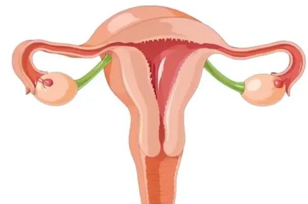
The normal size of uterus refers to the typical dimensions of the organ that supports pregnancy, basically a small, hollow muscle in the pelvis. Think of it like a flexible pouch that can stay compact or expand dramatically when needed. This sets up why even slight changes in size can matter.
The normal size of the uterus, described with a slight variation in phrasing, varies with age, childbirth history, and hormonal status. In most adults, it generally measures close to the size of a small pear, and shifts are common after pregnancies or during midlife. These patterns are well-documented, and they help explain why size ranges differ so much among individuals.
After routine checkups, when someone hears measurements that sound a bit larger or smaller than expected, they’re often just seeing the normal range of a uterus that adapts throughout life. Understanding what influences those changes makes it easier to follow the later discussion on causes, symptoms, and when evaluation is actually needed.
Average Uterine Size in Adults
(2.5–4 cm)
(2 cm)
(7.5 cm)
(5 cm)
(9–10 cm)
(6.5 cm)
(5–6 cm)
(4 cm)
For adult women who have never been pregnant, the average uterus is about 3 inches (7.5 cm) long, 2 inches (5 cm) wide, and 1 inch (2.5 cm) thick, weighing roughly 1.6–2 ounces (45–60 grams). That’s about the size and weight of a small pear. In women who have given birth, the uterus tends to be slightly larger—around 3.5 to 4 inches (9–10 cm) in length.
Health Support: This Vitamin K2 + D3 Complex is essential for bone density, cardiovascular health, and immune function. It’s a highly-rated formula for those looking to maintain optimal nutrient levels. You can find it on Amazon.
According to data from the American College of Obstetricians and Gynecologists, up to 70% of women experience minor uterine size variations without any medical concern. In most cases, these differences are due to hormones or childbirth history. ⧉
Uterine Size Through Life Stages
Childhood and Puberty
Before puberty, the uterus is very small—about 1 to 1.5 inches (2.5 to 4 cm) long. When estrogen levels rise during puberty, the uterus grows rapidly, preparing for menstruation and future fertility.
Reproductive Age
Between ages 18 and 45, the uterus maintains its adult size but temporarily enlarges slightly during menstrual cycles. This occurs because the endometrium (inner lining) thickens each month to prepare for a possible pregnancy.
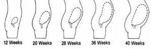
After Pregnancy
During pregnancy, the uterus grows incredibly—from the size of a pear to that of a watermelon. That’s roughly a 500-fold increase in volume. After childbirth, it shrinks back gradually over 6–8 weeks, a process known as uterine involution. However, it may remain 10–20% larger than before pregnancy.
Health Support: This high-absorption Magnesium Glycinate (200 mg) is gentle on the stomach and supports muscle relaxation, better sleep, and metabolic health. You can find this trusted formula on Amazon.
Menopause and Beyond
After menopause, the uterus often shrinks again due to the decline in estrogen. By age 60, it may measure only 2 to 2.5 inches (5–6 cm) long, roughly half its peak reproductive size.
How Doctors Measure Uterine Size
Understanding how the uterus is measured helps patients feel more comfortable during examinations. Each diagnostic method has its own purpose, accuracy, and comfort level. Let’s break down the most common ways doctors assess uterine size.
1. Ultrasound (Accuracy: 9/10)
Ultrasound is considered the gold standard for measuring uterine size because it’s safe, non-invasive, and provides immediate results. It uses high-frequency sound waves to create an image of the uterus, which is displayed on a monitor. There are two main types:
- Transabdominal ultrasound: A probe is moved across the lower abdomen with a gel that improves sound transmission. This method is more comfortable and used for general screening.
- Transvaginal ultrasound: A small probe is inserted into the vagina, providing a clearer image of the uterus and surrounding tissues. It’s more precise and often used for women with suspected fibroids, cysts, or abnormal bleeding.
The procedure takes about 15–30 minutes, doesn’t require anesthesia, and costs around $150–$300. Most women describe it as mildly uncomfortable but not painful. It’s typically used during routine gynecological exams, pregnancy assessments, or when symptoms like pelvic pain or irregular periods appear. ⧉
2. MRI Scan (Accuracy: 10/10)
MRI (Magnetic Resonance Imaging) is like the high-definition camera of medical diagnostics. It uses powerful magnets and radio waves to create a detailed 3D image of the uterus and surrounding organs. Unlike ultrasound, MRI can distinguish between different tissue types, making it invaluable for detecting fibroids, adenomyosis, or structural anomalies.
The process takes about 30–60 minutes, during which the patient lies inside a large scanner. It’s completely painless, though some may feel claustrophobic. Doctors often recommend MRI when ultrasound findings are unclear or when planning complex treatments. It’s the most accurate option, but also the most expensive—$700–$1,200 on average. ⧉
3. Pelvic Exam (Accuracy: 6/10)
A pelvic exam is the oldest and simplest method, performed during routine gynecological visits. The doctor uses gloved fingers and a speculum to feel the uterus’s size, position, and texture through the vaginal wall. Think of it as an initial physical check-up for the uterus—quick, low-cost, and often the first step before imaging tests.
Although it’s less precise than ultrasound or MRI, a skilled gynecologist can detect significant abnormalities, such as an enlarged or displaced uterus. The exam lasts just 5–10 minutes and costs about $100–$200. It’s slightly uncomfortable but safe and essential for preventive care. ⧉
Choosing the Right Diagnostic Method
The choice of method depends on symptoms, medical history, and doctor recommendations:
- Routine screening or checkups: Start with a pelvic exam.
- Unclear pain, abnormal bleeding, or suspected fibroids: Ultrasound is the go-to test.
- Complex cases or surgical planning: MRI provides the clearest and most detailed image.
According to Reyus Mammadli, medical consultant, “A woman’s comfort during diagnostics is just as important as accuracy. The key is to start with what’s least invasive and move forward only if needed.”
Factors That Influence Uterine Size
The uterus doesn’t exist in isolation—it responds to changes in hormones, health, and even lifestyle. Think of it as a “mirror” reflecting what’s happening throughout the body. Here’s a deeper look at the main factors that influence its size and health.
1. Hormonal Balance – The Master Regulator
Hormones are like the thermostat of the reproductive system. When estrogen and progesterone are balanced, the uterus stays within a healthy size range. But if estrogen rises too high (for example, due to obesity or certain medications), the uterus can enlarge. Low estrogen, such as after menopause, can cause it to shrink.
To maintain hormonal balance, women should focus on proper sleep, stress management, and a diet rich in whole grains, leafy greens, and omega-3s. Even mild stress can cause hormonal fluctuations that affect uterine health. As Reyus Mammadli often notes, “When hormones swing, the uterus follows.”
2. Pregnancy and Childbirth – Stretching with Purpose
During pregnancy, the uterus performs one of nature’s most astonishing transformations. It expands from the size of a pear to fit a full-term baby—sometimes weighing up to 15 times its original mass. After childbirth, it gradually returns to near-normal size but rarely to its pre-pregnancy dimensions.
Multiple pregnancies can lead to a slightly larger uterus permanently. It’s completely normal—like a balloon that’s been inflated and deflated several times. Gentle postpartum exercise, proper nutrition, and sufficient rest help the uterus recover effectively.
3. Medical Conditions – Hidden Influencers
Certain health problems can enlarge or shrink the uterus. Fibroids act like small “speed bumps” inside the uterine wall, pushing it outward. Adenomyosis makes the muscle thicker and heavier, while endometrial hyperplasia thickens the inner lining. ⧉
Regular checkups and ultrasounds are key. These conditions are often manageable if detected early. Ignoring prolonged pain or bleeding can allow the problem to grow silently for years.
4. Body Weight and Nutrition – The Daily Impact
Extra body fat can increase estrogen levels, subtly enlarging the uterus over time. Imagine estrogen as fertilizer—it helps tissue grow, but too much can cause overgrowth. On the other hand, extreme dieting or malnutrition can reduce hormone production, leading to uterine shrinkage.
Maintaining a balanced weight, eating iron-rich foods like spinach and beans, and staying hydrated all support healthy uterine function. Even walking 30 minutes a day can improve hormonal balance.
5. Genetics – Family Patterns
Some women are simply born with slightly larger or smaller uteruses. Genetic factors may determine the organ’s structure, elasticity, and even how it responds to hormones. If a woman’s mother or sisters have fibroids or other uterine conditions, she should be more proactive about monitoring her reproductive health through routine exams.
6. Age and Natural Changes
Age affects everything—including the uterus. In youth, it’s firm and muscular. With age and declining estrogen, it softens and reduces in size. This is a natural process, not a disease. Staying active and keeping bones and muscles strong through diet and exercise indirectly supports uterine health too.
In summary, uterine size is influenced by a mix of internal and external factors. While some—like genetics and aging—can’t be changed, others, such as hormone balance, nutrition, and body weight, are within your control. A healthy lifestyle truly supports a healthy uterus.
When Uterine Size Isn’t Normal
A uterus that is noticeably larger or smaller than the normal range can signal various health conditions. Let’s take a closer look at the main causes, using simple comparisons to make them easier to understand.
Fibroids – The Unwanted Houseguests
Fibroids are benign (noncancerous) muscle growths that develop inside or on the outer walls of the uterus. Imagine small balloons growing within the walls of a house—they push outward, making the house (the uterus) look bigger. Fibroids can vary in size from a pea to a grapefruit. About 1 in 3 women over 35 experience fibroids to some degree. Symptoms often include heavy bleeding, pelvic pressure, or bloating. Although noncancerous, large fibroids may require treatment through medication or minimally invasive surgery.
Adenomyosis – The Soft Wall Problem
Adenomyosis occurs when the tissue that normally lines the inside of the uterus starts growing into the uterine muscle itself. Think of it like tree roots growing into concrete—the structure becomes thicker and heavier. This condition can make the uterus feel enlarged and tender, and it often causes painful and heavy periods. It’s most common in women aged 35–50. Treatments include hormonal therapy or procedures that reduce the overgrown tissue.
Congenital Malformations – Born Different
Some women are born with a uterus that’s naturally smaller or shaped differently. A common example is the bicornuate uterus, shaped like a heart instead of a pear. These differences are usually harmless but can sometimes cause fertility issues or complications during pregnancy. With modern imaging and proper prenatal care, most women with these variations can still carry healthy pregnancies.
Hormonal Imbalance – The Size Controller
Just as plants need sunlight to grow, the uterus depends on estrogen and progesterone. When these hormones are out of balance—due to menopause, thyroid issues, or stress—the uterus may shrink or enlarge slightly. This change is usually temporary and reversible once hormone levels normalize.
If a woman experiences symptoms like pelvic pain, bloating, or unusually heavy periods, it’s time to check the uterine size through an ultrasound. Early detection can prevent minor issues from turning into major problems.
Real Case from the U.S.
A 39-year-old woman from Florida complained of prolonged periods and bloating. Her transvaginal ultrasound revealed her uterus measured 5.7 inches (14.5 cm) due to multiple fibroids. She underwent uterine artery embolization—a minimally invasive procedure that cuts off blood supply to fibroids. Within five months, her uterus returned to a healthy 3.8 inches (9.6 cm), and her symptoms resolved.
Editorial Advice
Reyus Mammadli, a medical consultant, emphasizes that a slight difference in uterine size is perfectly normal. What matters more are the symptoms and rate of change. Women should schedule annual pelvic exams and ultrasounds every 2–3 years, especially after 35.
Maintaining a balanced diet rich in omega-3 fatty acids, vitamin E, and iron supports uterine tissue health. Foods like salmon, lentils, and spinach are beneficial. Hydration and maintaining a healthy weight also help regulate hormones naturally.
Finally, listen to your body. Persistent pain, irregular bleeding, or a feeling of pelvic fullness should never be ignored. Early detection through imaging and lab tests can prevent serious conditions such as fibroids or endometrial hyperplasia from progressing. As Reyus Mammadli advises: “A healthy uterus is often silent—when it starts ‘speaking,’ it’s time to listen carefully.”