A rare yet severe side effect of heparin, a medication used to prevent blood clots, is skin necrosis. This condition, in which skin sores and tissue death occur at the site of injection, can lead to serious health complications.
Causes of Heparin-induced skin necrosis
Skin necrosis caused by heparin usually happens because of an immune response. This response can be a result of heparin-induced thrombocytopenia (HIT), a condition where heparin causes the production of abnormal antibodies that activate platelets. This can lead to clot formation and, ironically, increase the risk of both bleeding and blood clots.
- Immune reaction: In some individuals, heparin can trigger an immune response leading to the formation of antibodies, which in turn can cause skin necrosis.
- Low protein C levels: Heparin can reduce the levels of protein C, a natural anticoagulant in the body. Low levels of protein C can increase the risk of skin necrosis.
- Heparin sensitivity: Some people may have a specific sensitivity or allergy to heparin, which can manifest as skin necrosis when the medication is administered.
- Underlying conditions: Patients with conditions such as cancer, sepsis, or antiphospholipid syndrome may be at a higher risk of developing heparin-induced skin necrosis.
- Dosage-related: High doses of heparin or rapid administration can sometimes overwhelm the body’s natural mechanisms, leading to skin necrosis in susceptible individuals.
- Delayed onset: Skin necrosis due to heparin administration may not always occur immediately; it can sometimes develop days to weeks after starting therapy.
- Arterial thrombosis: Heparin-induced skin necrosis is more commonly associated with arterial thrombosis, where blood flow to the tissues is compromised, leading to skin damage.
Symptoms
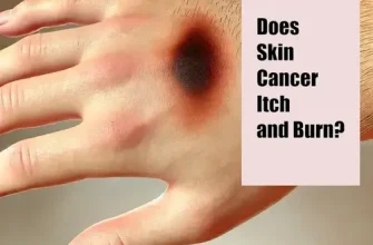
The primary symptom is the appearance of painful, reddened skin lesions at sites where heparin has been injected. Over time, these lesions can evolve into areas of necrosis (tissue death), characterized by darkening of the skin which may become black, indicating full-thickness skin death.
When it comes to heparin-induced skin necrosis, being aware of the symptoms is crucial for prompt diagnosis and treatment. Here are some key signs to watch out for:
- Localized Pain: Individuals may experience severe pain, tenderness, or a burning sensation at the site where heparin was administered. This pain may gradually worsen over time.
- Skin Discoloration: One of the earliest signs of heparin-induced skin necrosis is the development of a purple or reddish discoloration at the injection site. This may progress to a dark, bruise-like appearance.
- Skin Necrosis: As the condition advances, the skin at the injection site may begin to feel firm, followed by the development of a deep, ulcerated wound. This necrotic tissue is a critical indicator of the severity of the condition.
- Blistering or Ulceration: Heparin-induced skin necrosis can lead to the formation of blisters or ulcers at the injection site. These lesions may be painful, ooze fluid, or become crusted over time.
- Necrotic Lesions: In severe cases, the affected skin may undergo necrosis, turning black and eventually sloughing off. This indicates extensive tissue damage and requires immediate medical attention.
- Systemic Symptoms: Some individuals with heparin-induced skin necrosis may also experience systemic symptoms such as fever, chills, malaise, or fatigue. These symptoms could be a sign of a more widespread inflammatory response in the body.
- Delayed Onset: It is important to note that symptoms of heparin-induced skin necrosis may not appear immediately after the administration of heparin but can manifest hours to days later. Therefore, vigilance is key in monitoring for any unusual skin changes post-heparin therapy.
Diagnosis
Diagnosis of heparin-induced skin necrosis is primarily clinical, based on the patient’s history, presentation, and timing of symptoms in relation to heparin administration. Laboratory studies are important to confirm the presence of HIT antibodies or decreased platelet counts, helping to differentiate this condition from other causes of skin necrosis.
| Diagnostic Step | Description |
|---|---|
| Clinical Exam | Assessment of skin lesions and medical history regarding heparin exposure |
| Lab Tests | Platelet count monitoring, HIT antibodies testing |
| Imaging | To assess for deep vein thrombosis if HIT is confirmed |
Treatment Options for Heparin-induced Skin Necrosis
Immediate discontinuation of heparin is the primary treatment for heparin-induced skin necrosis. Alternative anticoagulants, such as direct thrombin inhibitors or factor Xa inhibitors, may be used instead. In severe cases, surgical intervention might be necessary to remove necrotic tissue and to prevent further complications, such as infection.
Here are the treatment options available:
| 1. Discontinue Heparin Therapy |
|---|
| The first step in managing heparin-induced skin necrosis is to stop the administration of heparin. This helps prevent the progression of skin necrosis and allows for the initiation of alternative therapies. |
| 2. Topical Treatments |
|---|
| Applying topical treatments such as corticosteroids or heparinoid creams can help reduce inflammation and promote healing of the affected skin. These creams can be applied directly to the necrotic areas several times a day. |
| 3. Wound Care |
|---|
| Proper wound care is crucial in managing heparin-induced skin necrosis. This includes keeping the affected area clean and dry, as well as dressing the wounds with sterile bandages to prevent infection. |
| 4. Hyperbaric Oxygen Therapy (HBOT) |
|---|
| HBOT involves breathing pure oxygen in a pressurized room or chamber. This therapy can improve oxygen delivery to the affected tissues, promote healing, and reduce skin necrosis. |
| 5. Surgical Interventions |
|---|
| In severe cases of heparin-induced skin necrosis, surgical interventions such as debridement (removal of dead tissue) or skin grafting may be necessary to promote wound healing and prevent complications. |
| 6. Anticoagulant Reversal Agents |
|---|
| If heparin-induced skin necrosis is associated with heparin-induced thrombocytopenia (HIT), administration of anticoagulant reversal agents such as protamine sulfate may be required to counteract the effects of heparin. |
| 7. Consultation with Specialists |
|---|
| Seeking consultation with dermatologists, wound care specialists, or hematologists can provide tailored treatment plans and optimize the management of heparin-induced skin necrosis. |
Early recognition and prompt treatment of heparin-induced skin necrosis are vital in preventing complications and promoting skin healing. It is essential to work closely with healthcare providers to determine the most appropriate treatment approach for individual cases.
How to Avoid
To avoid heparin-induced skin necrosis, it is crucial to monitor patients closely for any signs of adverse reactions to heparin, especially those receiving high doses or with previous history of HIT. Choosing alternative anticoagulants in high-risk patients may also reduce the risk.
Living with Heparin-Induced Skin Necrosis
Managing heparin-induced skin necrosis involves close medical monitoring and supportive care:
- Regular follow-up appointments to monitor the healing of skin lesions and overall health.
- Education about recognizing symptoms of complications, such as signs of infection or deep vein thrombosis.
- Psychological support to cope with the distress associated with disfiguring skin lesions.
- Discussion with healthcare providers about future anticoagulation management to prevent recurrence.
In conclusion, heparin-induced skin necrosis is a complex condition requiring timely intervention. Early diagnosis, cessation of heparin, appropriate alternative anticoagulation, and supportive care remain the cornerstones of managing this serious complication.

